
Fig 7.
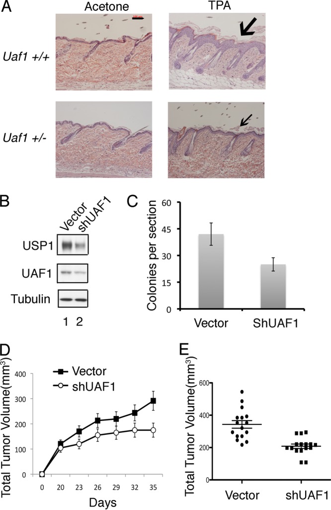
Uaf1 deficiency causes decreased cell proliferation and inhibits tumorigenesis. (A) Uaf1+/− mice have decreased TPA-induced epithelial proliferation. Six-week-old Uaf1+/− and Uaf1+/+ mice were exposed topically to TPA for 4 weeks. The mice were sacrificed, and the skin histology was evaluated by microscopy. H&E staining of tissue sections of skin (representative analysis; n = 4 for each genotype) is shown. The scale bar represents 100 μm, and the arrows indicate epithelial layers. (B to E) UAF1 knockdown in lung cancer cells suppresses growth in soft agar and reduces tumorigenesis in nude mice. (B) The cDNA encoding a UAF1 shRNA or shScramble (Vector) was stably expressed in the RAS-driven human lung adenocarcinoma epithelial cell line A549. The cells were subjected to growth assay or analyzed for tumorigenicity in nude mice. Immunoblots of the A549 cell lysates are shown. (C) Usp1 antibody against the N-terminal epitope of Usp1 was used for immunoblots. A549 cells were plated in soft agar, and transformed foci were counted (at ×100 magnification) after 2 weeks in culture. The data are means ± standard errors of three independent experiments. Each experiment was performed in triplicate plates (P < 0.001; t test). (D and E) Tumorigenicity of A549 cells in xenograft mouse models. UAF1 shRNA- or shScramble-transfected A549 cells were injected subcutaneously as xenografts in nude mice (two-site injections), and every 3 days the tumor volume was measured. The data represent the average tumor size for 10 mice in each group (more than 10 tumors, due to the two-site injections; P < 0.001). Quantification of the tumor volume of the xenografts at day 35 is shown in panel E. The data are means ± standard errors.