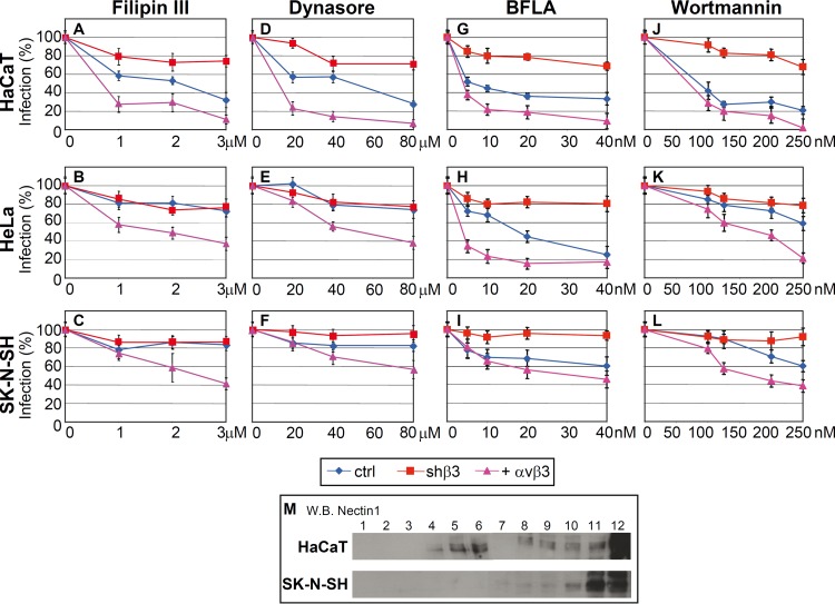
Fig 4.
(A to L) Effect of inhibitors on infection of R8102 into HaCaT, HeLa, and SK-N-SH cells. Cells were mock silenced (ctrl), had β3 integrin silenced (shβ3), or transiently overexpressed αvβ3 integrin (+ αvβ3). The stock solutions of filipin III (2.5 mM), dynasore (100 mM), bafilomycin A (BFLA) (160 mM), and wortmannin (2 M) (all from Sigma-Aldrich) in dimethyl sulfoxide were stored at −20°C. Cells were exposed to the indicated amounts of inhibitors for 1 h at 37°C and then infected with R8102 (3 PFU/cell) for 90 min in the presence of inhibitors (18). The viral inoculum was removed, and the cells were overlaid with medium containing inhibitors for 6 to 8 h. For filipin III and dynasore, cells were preincubated with the compounds at 37°C for 30 min and 60 min, respectively, and infected for 30 min (30 PFU/cell) in the same medium. Viral inoculum was removed; infected cells were overlaid without inhibitor and harvested 6 to 8 h after infection. Infection of R8102 was quantified by means of o-nitrophenyl-β-d-galactopyranoside; each point represents the average of triplicates ± standard deviation. (M) Flotation of membranes from HaCaT and SK-N-SH cells. Cells were solubilized in TNE buffer (10 mM Tris-HCl, 150 mM NaCl, 5 mM EDTA, 1% Triton X-100, 0.036 mg/ml each of the protease inhibitors Nα-p-tosyl-l-lysine-chloromethyl ketone [TLCK; Sigma-Aldrich] and N-tosyl-l-phenylalanine-chloromethyl ketone [TPCK; Sigma-Aldrich]) and layered at the bottom of a 5-35-42% sucrose gradient as detailed previously (39). After centrifugation at 34,000 rpm for 20 h at 4°C in a SW41 swing-out rotor, 12 fractions were collected from the top. Aliquots from each fraction were subjected to sodium dodecyl sulfate-polyacrylamide gel electrophoresis. Nectin-1 was detected by Western blotting (WB) with CK6 monoclonal antibody (Santa Cruz Biotechnologies) followed by peroxidase-conjugated anti-mouse antibody and enhanced-advance chemiluminescence (ECL-Advance kit; GE Healthcare). Numbers above the lanes indicate the numbers of the fractions. The part of the image relative to fractions 1 to 6 was exposed for 3 min; the part of the image relative to fractions 7 to 12 was exposed for 30 s.

