
Fig 1.
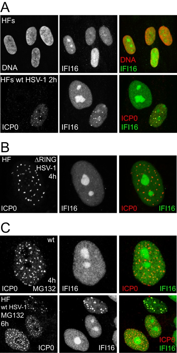
Localization of IFI16 in uninfected and infected HFs. (A) Upper row, Uninfected HFs were stained for IFI16 and DNA (Topro-3); lower row, HFs were infected with wt HSV-1 at an MOI of 4 and stained 2 h later for IFI16 (Abcam ab55328) and ICP0. (B) HFs were infected with HSV-1 expressing a RING finger deletion mutant ICP0 at an MOI of 4 and stained 4 h later for IFI16 and ICP0. (C) HFs were infected with wt HSV-1 at an MOI of 4 in the presence of 3 μM MG132 and stained 4 h and 6 h later for IFI16 and ICP0.