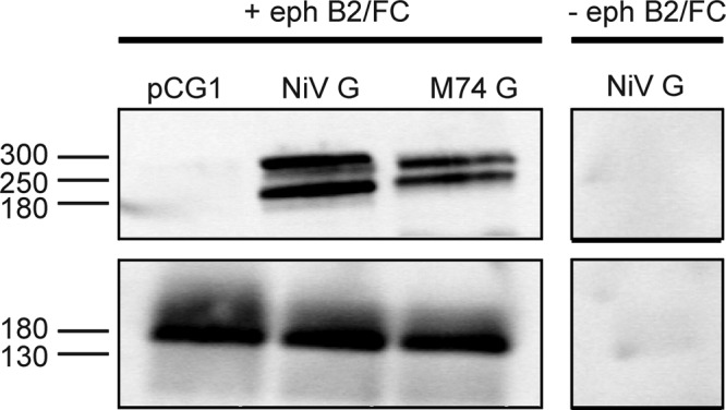
Fig 3.

Interaction of the G proteins of M74 and NiV with ephrin B2. The FLAG-tagged G proteins of NiV and M74 were expressed in BHK-21 cells. At 24 h p.t., cells were lysed in NP-40 lysis buffer and mixed with protein A-Sepharose (Sigma-Aldrich) preloaded with soluble Fc-tagged ephrin B2 (+ eph; R&D Systems). − eph, control. After several washings, bound G proteins were released from the beads by boiling in nonreducing sample buffer and analyzed by Western blotting. G proteins were visualized by immunostaining (upper panels) using antibodies against the FLAG tag (Sigma-Aldrich). The presence of ephrin B2 was shown by immunostaining (lower panels) using antibodies against the Fc tag (Dako). Molecular mass markers (in kilodaltons) are indicated on the left.
