
Fig 8.
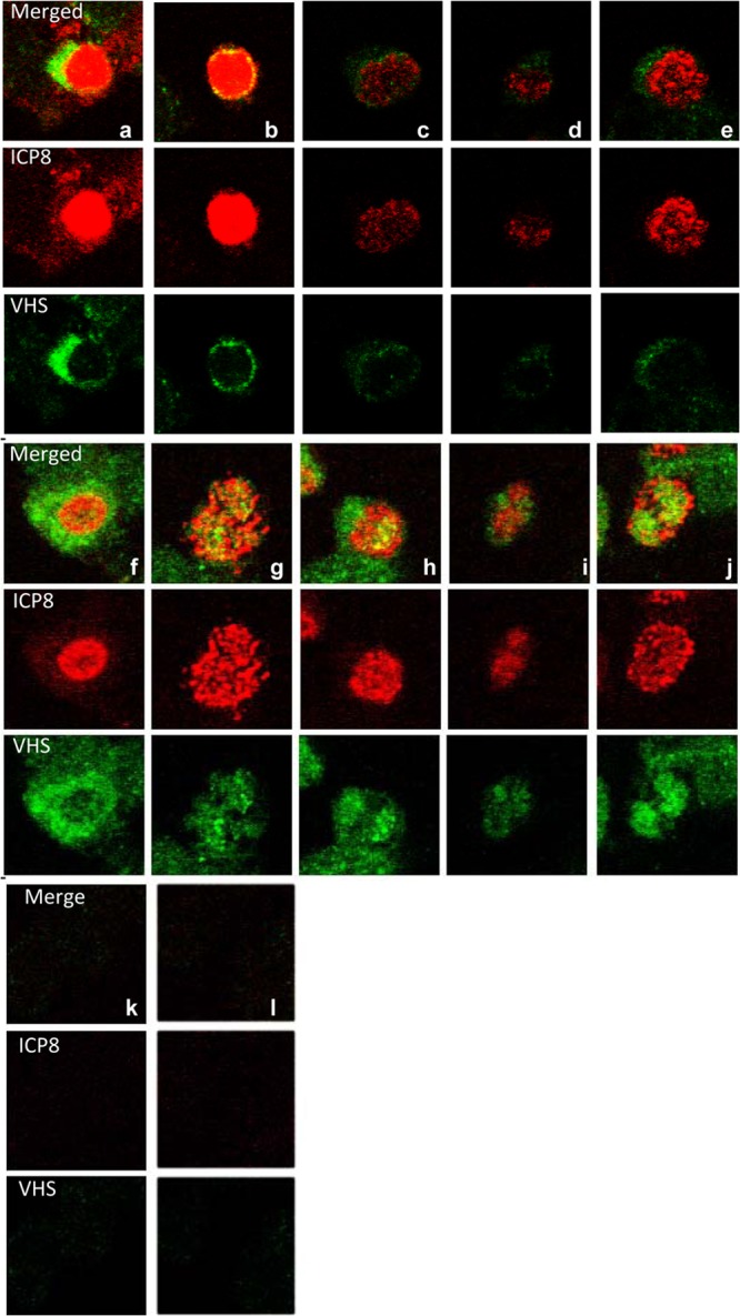
Wild-type VHS shuttles between the nucleus and cytoplasm. HEp-2 cells were mock infected or exposed (20 PFU/cell) to HSV-1(WT-BAC) virus for 1 h. The inocula were then replaced with fresh complete medium with or without leptomycin B (10 μM). The cells were fixed at 2 h after exposure to the virus and fixed and reacted with antibodies to VHS and ICP8. The images were collected with the aid of a Zeiss confocal microscope.