
Fig 4.
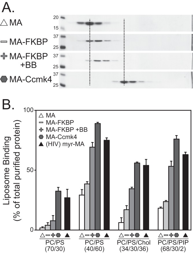
Flotation analysis of purified RSV MA multimers. (A) Velocity sedimentation of RSV MA, MA-FKBP, MA-FKBP +BB, and MA-Ccmk4 proteins. Molecular masses (in kDa) are indicated for the MA and MA-FKBP gels; all other gels were run identically. Dotted lines are added to more easily show the shift of MA monomers to dimer and hexamer. (B) Percentages of total protein found associated with four liposome types. Symbols indicate protein types as for Fig. 2. All MA proteins are RSV except HIV-1 myr-MA, which is indicated by the black triangle. Error bars represent standard deviations from the means.