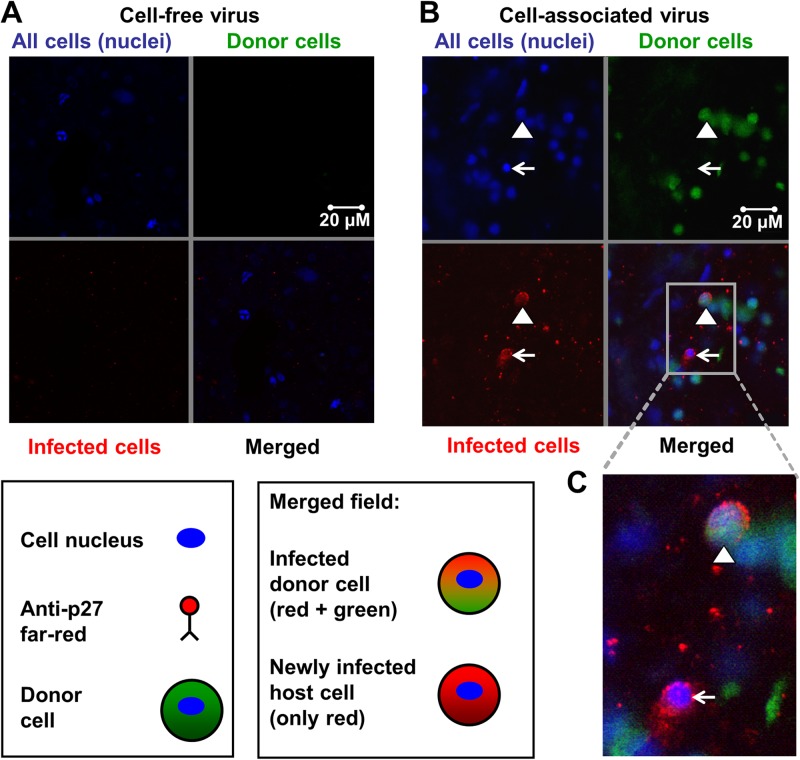
Fig 2.
SIVmac251-infected donor cells initiated host cell infections. One milliliter of plasma (2.4 × 10³ TCID50/ml) or 1 × 10⁶ PBMCs (18 TCID50/ml) from an SIV-infected monkey stained with CellTracker Green were added atop sealed colon explants obtained from SIV-naive rhesus macaque for 48 h. After viral exposure, organ explants were washed, fixed, and stained using whole-mount immunofluorescence. (A, B) DAPI stained cell nuclei blue (upper left quadrants), CellTracker Green stained donor cells green (upper right quadrants), and anti-p27 Far Red stained SIV p27 protein (SIV capsid) red (lower left quadrants). The lower right quadrants show the merged fields. For each of 2 independent experiments performed in triplicate, the entire tissue specimens (2 by 2 mm) were scanned by two independent investigators. (A) Representative confocal images of explants exposed to plasma containing cell-free SIVmac251. No infected cells were identified in the merged field after addition of free SIVmac251 atop sealed colon tissue. (B) Representative confocal images of explants exposed to PBMCs from an SIVmac251-infected monkey. The triangle points to an infected donor cell (green cytoplasm, red viral protein, blue nucleus), and the arrow points to a newly infected host cell (only red and blue staining). (C) High-powered view of the merged field.

