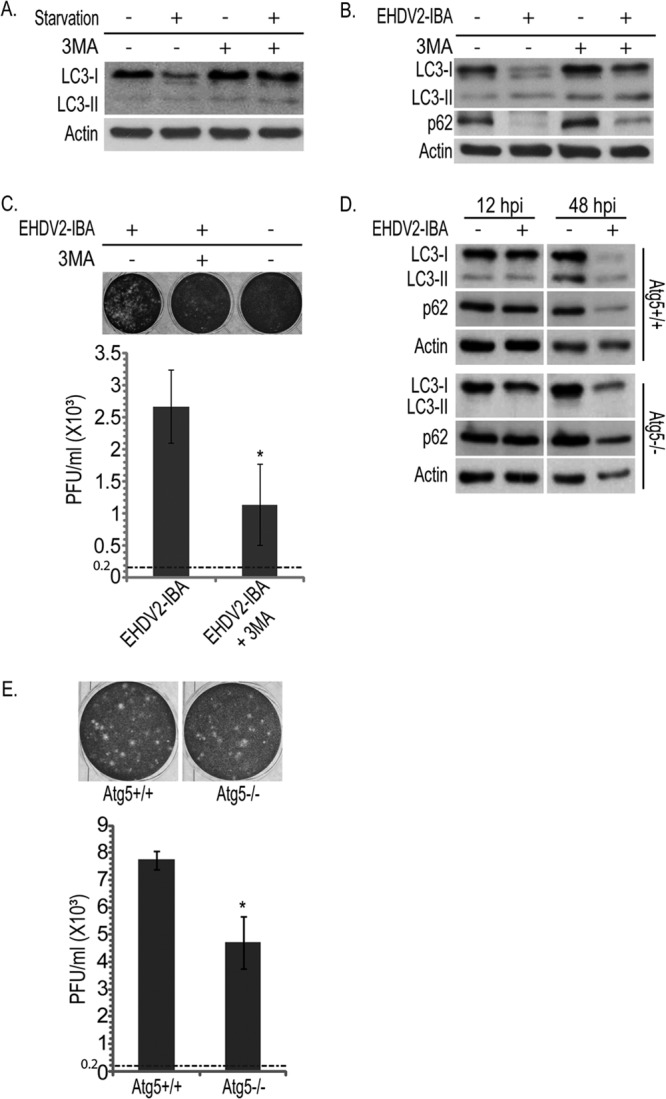
Fig 5.

Inhibition of autophagy attenuates the production of infectious particles. OK cells at 24 h postplating were starved or given EBSS (3 h) and treated with 3MA (10 mM for 3 h) or left untreated. Cells were lysed, separated by 12% SDS-PAGE, blotted, and probed with anti-LC3 or antiactin antibodies. (B) Representative immunoblot of lysates of OK cells infected or not with EHDV2-IBA (36 hpi) and treated or not with 3MA (10 mM for 36 h), separated by 12% SDS-PAGE, and probed with the indicated antibodies. (C) Plaque assay analysis of the titer of infectious virions under untreated or 3MA treatment conditions. The upper panels show representative wells displaying plaques formed upon infection of Vero cells with different dilutions of the lysates of OK cells (uninfected or infected with EHDV2-IBA; 36 hpi), untreated or treated with 3MA (10 mM; 36 h). The graph in the lower panel depicts the titer (PFU/ml) produced in infected OK cells (untreated or treated with 3MA) at 36 hpi. The titer of the inoculum was 0.2 × 10³ PFU/ml and is represented by the dotted line. *, P < 0.05. (D) Atg5+/+ or Atg5−/− MEFs at 24 h postplating were left uninfected or infected with EHDV2-IBA for 12 or 48 h. Cells were lysed, separated by 12% SDS-PAGE, blotted, and probed with antibodies against LC3, p62, or actin. (E) Plaque assay analysis of the titer of infectious virions generated in infected Atg5+/+ or Atg5−/− MEFs. Sample processing and data presentation are as described for panel C.
