
Fig 1.
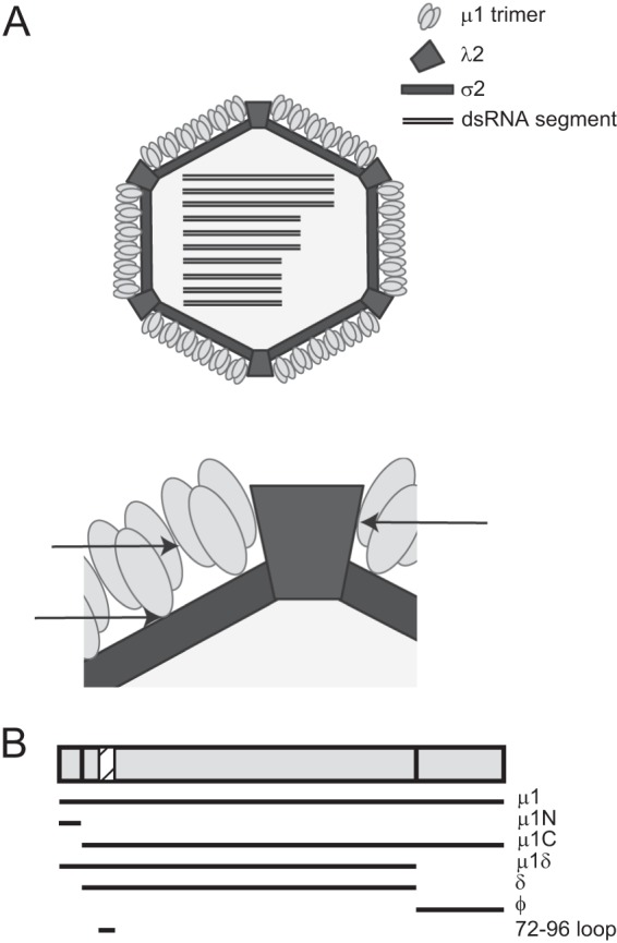
Schematic of the μ1 protein and its interactions within the virus particle. (A) A broad (top) and zoomed-in (bottom) schematic of the cutaway view of an ISVP showing the arrangement of the μ1 protein surrounding the core. Arrows indicate regions of interaction between μ1 trimers and between trimers and the core that could be mediated by the 72-96 loop. (B) The various fragments of μ1 generated during cell entry of reovirus are shown. The location of the μ1 72-96 loop is shown as a hatched box.