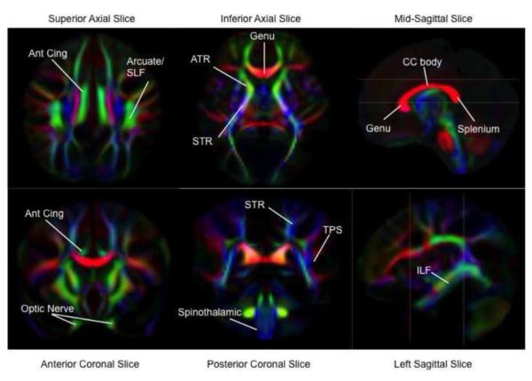
Figure 2.
DTI Fractional Anisotropy (FA) Maps of White Matter Tracts. FA maps are color-coded by local fiber direction. The colors, red, green, and blue represent white matter fibers running along the right-left, anterior-posterior, and superior-inferior axes, respectively. Locations of white matter tracts are assigned on the color maps. Reference lines in sagittal images indicate respective locations of axial and coronal images. Temporal-parietal segment (TPS), inferior longitudinal fasiculus (ILF), body of the corpus callosum (CC Body), anterior cingulum (Ant. Cing), anterior thalamic radiations (ATR) part of the anterior limb of the internal capsule, superior thalamic radiations (STR) part of the posterior limb of the internal capsule.

