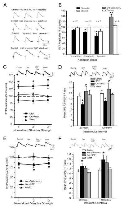
Figure 1.
(A) Nociceptin (Noc) dose-dependently decreases gamma-aminobutyric acid (GABA)ergic transmission in central nucleus of the amygdala (CeA) neurons. Representative recordings of evoked inhibitory postsynaptic potentials (IPSPs) in a CeA neuron from a naive rat recorded before, during Noc or corticotropin-releasing factor (CRF; 20 min), and washout (30 min). (B) Histograms representing percent decrease in mean ± SEM evoked IPSP amplitudes. *p < .05 and **p < .01. (C) Top panel: representative recordings of evoked IPSPs. Bottom panel: CRF increases mean IPSP amplitudes and subsequent application of Noc diminishes the CRF-induced effect. (D) Top panel: representative recordings of evoked 50-msec paired-pulse IPSPs in a CeA neuron. Bottom panel: CRF significantly decreases the 50- and 100-msec paired-pulse facilitation ratio of IPSPs. #p < .05 indicates significance between CRF alone and concurrent application of CRF and Noc. (E) Noc prevents CRF-induced enhancement of GABAergic transmission in CeA neurons. Top panel: Representative recordings of evoked IPSPs. Bottom panel: superfusion of 500 nmol/mL Noc significantly decreases mean IPSPs and prevents the CRF-induced enhancement. (F) Top panel: representative recordings of evoked 50-msec paired-pulse IPSPs. Bottom panel: Noc significantly increases the 50- and 100-msec paired-pulse facilitation ratio of IPSPs and blocks the CRF-induced decrease in paired-pulse facilitation ratio (p < .05).

