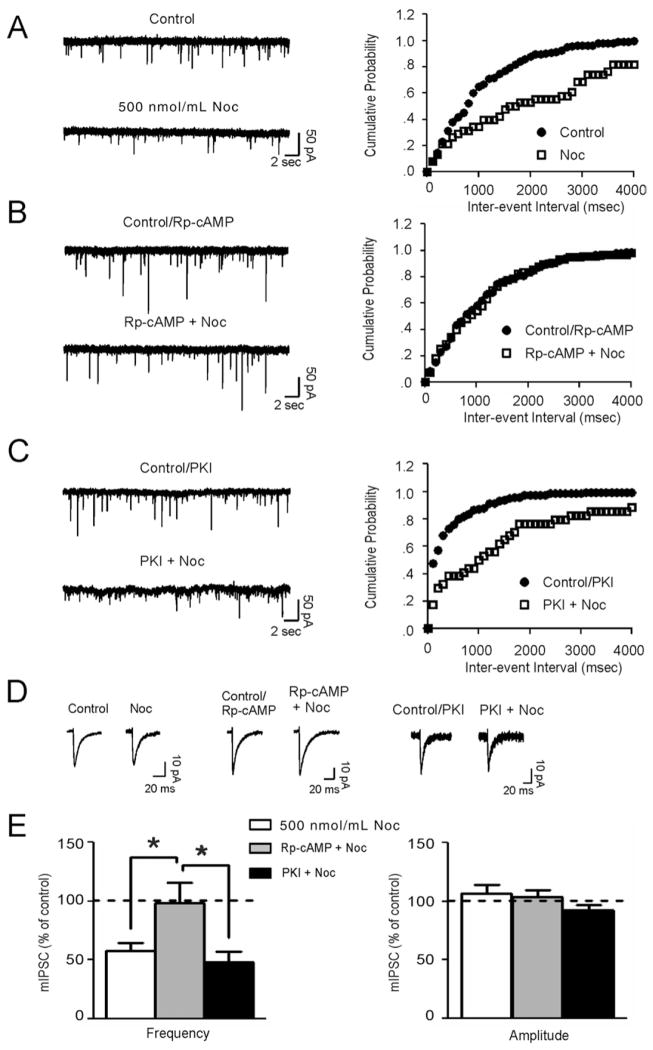
Figure 5.
Nociceptin (Noc) decreases gamma-aminobutyric acid release via a protein kinase A (PKA)-dependent pathway. (A) Left panel: representative miniature inhibitory postsynaptic currents (mIPSCs) from a central nucleus of the amygdala (CeA) neuron before and during 500 nmol/mL Noc application. Right panel: a rightward shift in the cumulative frequency plot for the neuron in Figure 5A indicating a longer interevent interval during the application of Noc. (B) Left panel: representative mIP-SCs from a CeA neuron before and during application of nociceptin in the presence of Rp-cAMP. Pretreatment with Rp-cAMP prevented Noc from decreasing mIPSC frequency. Right panel: cumulative frequency plot for the CeA neuron in Figure 5C. (C) Left panel: representative mIPSCs before and during application of nociceptin with 5 μmol/mL PKI. Right panel: a rightward shift in the cumulative frequency plot for the CeA neuron in Figure 5E indicates a longer interevent interval during the application of nociceptin with PKI in the internal solution. (D) Scaled average mIPSCs from the traces depicted in Figures 5A–C. (E) Histograms depicting the average change in mIPSC frequency (left panel) and amplitude (right panel) with nociceptin alone, nociceptin in the presence of Rp-cAMP, and nociceptin with PKI in the internal solution (between subjects one-way analysis of variance: [F(2,13) = 5.55; *p < .05]).

