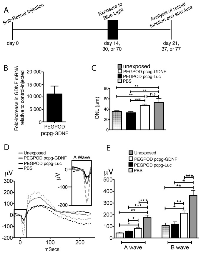
Figure 3. Injection of PEGPOD pcpg-GDNF NPs 14 days prior to blue light exposure provides complete structural and significant functional rescue of murine retina.
(A) Schema depicting the time course of experiments described in this study (B) QRT-PCR analysis of GDNF mRNA at 14 days post injection of eyes with PEGPOD pcpg-GDNF NPs shows an 11,200 fold increase in GDNF signal relative to eyes injected with PBS. n=4 (C) Average ONL thickness of the superior hemisphere within 500μm of the optic nerve head 7 days following exposure to blue light. ONL thickness, measured at 21 days post injection, shows a significant increase in ONL thickness of retinas injected with PEGPOD pcpg-GDNF NPs relative to PBS and PEGPOD pcpg-Luc NP injected (p<0.01, p<0.0005, respectively). Unexposed eyes were injected with PBS 21 days prior to analysis and unexposed to blue light. PBS, n=5; PEGPOD pcpg-GDNF, n=13; PEGPOD pcpg-Luc, n=10. Data is shown as mean+SEM. (D) Average Erg tracings of eyes injected with PEGPOD pcpg-GDNF NPs, PEGPOD pcpg-Luc NPS, or PBS 14 days prior to light exposure were recorded at 7 days following exposure to blue light (21 days post injection). An amplified image of the boxed region of the ERG (A-wave) is also shown. Unexposed eyes were injected 21 days prior to analysis but unexposed to blue light. (E) Average A- and B-wave amplitudes of eyes injected with PEGPOD pcpg-GDNF NPs, PEGPOD pcpg-Luc NPs, or PBS 14 days prior to light exposure. PEGPOD pcpg-GDNF NP injected eyes show significantly increased A- and B-wave amplitudes relative to both PEGPOD pcpg-Luc NP injected (p<0.05, p<0.01 for A-wave and B-wave amplitudes, respectively) and PBS injected (p<0.05 for both A- and B-wave amplitudes) eyes. Data is shown as mean+SEM. ONL, outer nuclear layer; n.s., not significant.

