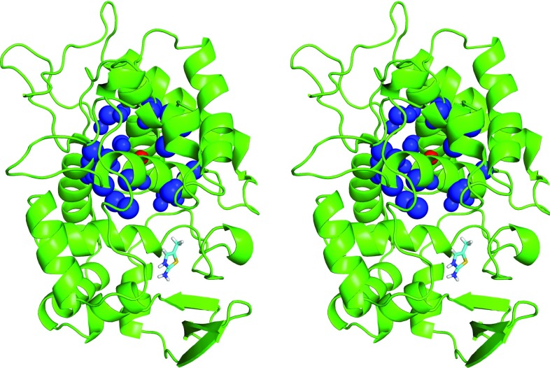
Figure 1.
Protein-ligand test system. Stereo view of the charged ligand 2-amino-5-methylthiazole (stick representation) bound to the engineered binding site of yeast cytochrome c peroxidase (CCP W191G “Gateless”47). The atomic sites used in the system with protein net charge +9 e for introducing an additional quasi-isotropic quadrupole moment (system net9quad) are also shown, namely, a central point charge of magnitude −80 e (red sphere) and 36 peripheral sites of total charge +80 e within a distance range of 0.81–0.85 nm (blue spheres). Note that the ligand binding mode is the one used in the present simulations, originally chosen based on the experimental binding mode of the same ligand to a related mutant protein,89 and does not exactly correspond to the experimentally inferred binding mode for the CCP W191G “Gateless” mutant.48

