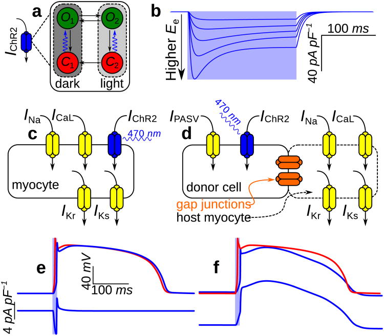
Figure 2. Modelling ChR2 properties and delivery modes at the cell level.
(a) Four-state Markov model for IChR2 with dark- and light-adapted photocycle branches associated with different peak conductances; each branch comprises closed- (red) and open-channel (green) states. Channel opening rates (blue arrows) are directly proportional to the irradiance (Ee) of light absorbed by ChR2 and open channels approach an Ee-dependent equilibrium between open states O1 and O2; all other transitions are purely time-dependent. (b) IChR2 in response to illumination (pale blue background; top to bottom: Ee = 0.4, 1, 2, 4, and 10mW mm−2) with Vm clamped to —85.6mV. Photoevoked ChR2 current has well-defined transient and steady-state phases resulting from the transition from full dark adaptation to an equilibrium between the two operating modes. The IChR2 model qualitatively reproduces experimental records from whole-cell patch-clamp recordings in a stable HEK-ChR2 cell line21, as shown in Supplementary Fig. S1. (c&d) Schematics for modelling gene delivery (GD) and cell delivery (CD) of ChR2; generic ionic currents are shown in myocytes. (e) Optically- (blue) and electrically-evoked (red) action potentials (APs) and underlying IChR2 in a myocyte with gene-delivered ChR2. Illumination at 2× threshold (Ee = 0.468mW mm−2 over 10ms) elicited depolarising IChR2 (bottom), which triggered an optically-evoked AP. (f) Response to illumination in an inexcitable ChR2-rich donor cell (CD; dashed blue) coupled to a normal myocyte by a 500 MΩ resistance (solid blue); a photoevoked AP in the latter cell is compared to an electrically-evoked AP (red). Light delivered to the donor cell was at 2× threshold for optically eliciting an AP in the myocyte (Ee = 0.652mW mm_2 over 10ms). During the AP plateau phase, myocyte Vm (≈ 2.77mV) was between the plateau Vm of the electrically-induced AP (≈ 15.3mV) and the donor cell resting level (−40mV).

