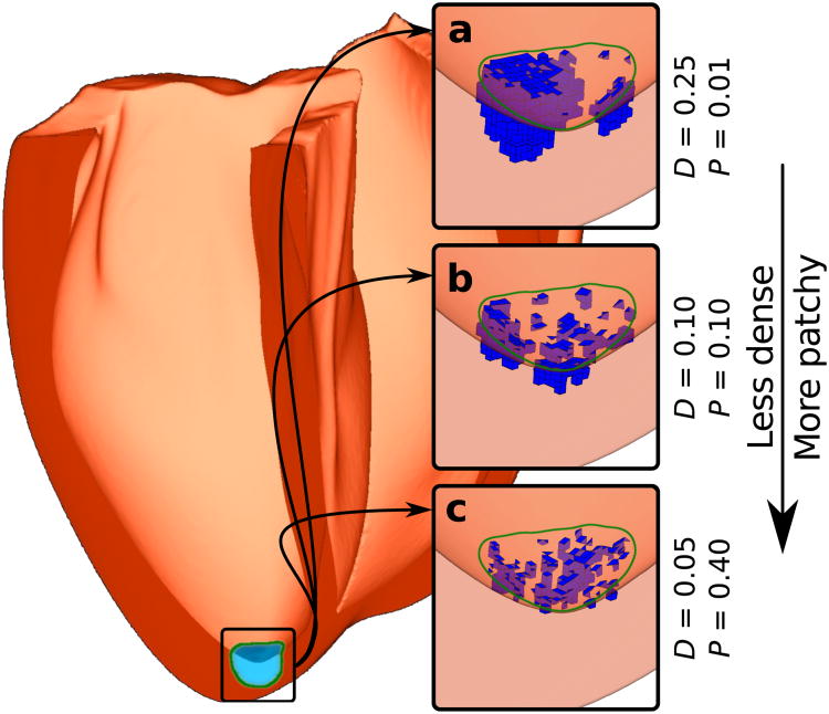
Figure 3. Modelling spatial distribution of light-sensitive cells at the tissue level.
Human ventricular model with a photosensitisation target (green boundary; hemispherical, 1 cm diameter) near the LV apex. (a-c) Results of applying the light-sensitive cell distribution algorithm to populate the target region with framework-generated ChR2-expressing clusters (blue) for three combinations of the parameters D (density) and P (patchiness).

