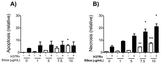
Figure 6. Pre-exposure to CS increased necrosis of Mtb-infected macrophages.
THP-1 macrophages were exposed to CS at concentrations of 1, 5 and 10 μg/ml for 24 h, then the macrophages were infected with Mtb. Apoptosis (A) or necrosis (B) was evaluated 24 h after Mtb-infection. Staurosporine (3 μM) was used as a positive control. Data is re_’¡. Bars indicate mean ± SD. *P<0.05. ANOVA and Dunnett’s post-hoc test compared to unexposed macrophages.

