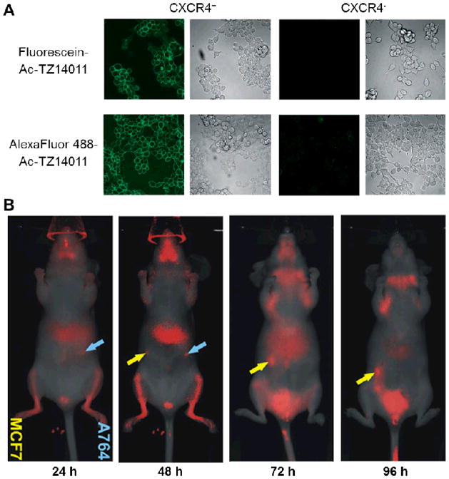
Fig. 1.
Fluorescence imaging of CXCR4. A. Confocal microscopy imaging of CXCR4+ and CXCR4− cells using fluorescein- or AlexaFluor 488-labelled Ac-TZ14011. B. Serial in vivo imaging of mice implanted with MCF7 (yellow arrows) and A764 (cyan arrows) tumors after administration of IRDye 800CW-labelled CXCL12. Adapted from [46, 54].

