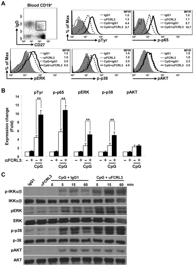
Figure 5. FCRL3 augments CpG-induced NF-κB and MAPK pathway activation.
(A) CD19+ blood B cells purified by negative selection were cultured overnight in the presence or absence of CpG. Cells harvested the next day were stained for CD19, IgD, and CD27 as well as biotinylated F(ab′)2 anti-FCRL3 or an IgG1 control before SA ligation and stimulation for 20 minutes at 37°C. Cells were immediately fixed, permeabilized, and intracellularly labeled with Abs specific for pTyr, p-p65, pERK1/2, p-p38, pAKT or respective conjugated control Abs. The dot plot shows gating of the CD19+CD27+IgD+ population from which staining in the histograms is derived. MFIR values resulting from the indicated stimulation conditions are shown in the histograms. The fold change of the MFIR relative to the unstimulated control is quantified in (B) using the mean MFIR ± SD and is representative of experiments from three different donors. ** P < 0.001 vs CpG plus the IgG1 control by two-tailed t-test. (C) SUDHL5 B cells were treated as in Fig. 2 with the specified stimuli. At the indicated time intervals, cells were lysed and immunoblotted with the listed phospho- or protein-specific Abs to ensure equal loading. One representative of three independent experiments is shown.

