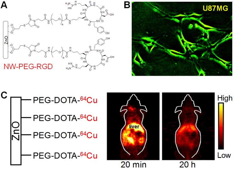
Fig. (3).
Targeted optical imaging with green fluorescent ZnO nanowires (NWs). A. A schematic structure of RGD peptide conjugated ZnO NWs. PEG denotes polyethylene glycol. B. Fluorescence imaging of integrin αvβ3 on U87MG human glioblastoma cells with NW-PEG-RGD. Magnification: 200×. C. Representatives positron emission tomography images of 64Cu-labeled non-targeted ZnO NWs at 20 min and 20 h postinjection into female Balb/c mice. Adapted from [44].

