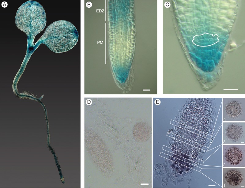
Fig. 1.
Spatial expression of Mob1A in arabidopsis seedlings. (A–C) GUS staining of 3- to 4-day-old pMob1A::GUS-Mob1A seedlings. In (B), the root proximal meristem (PM) and the beginning of the elongation–differentiation zone (EDZ) are labelled and indicated by white bars. In (C), the root stem cell niche is outlined in white. (D and E) In situ hybridization of specific sense (D) and antisense (E) Mob1A probes in longitudinal root sections. For (E) every second or every third 7 µm cross-section from the columella tissue up to the meristem is shown: (1) columella cells, (2) stem cell niche and (3 and 4) distal meristem cells. Scale bars are 10 µm in B and C, and 50 µm in D and E.

