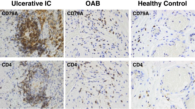
Fig. 1.
B- and T-lymphocyte staining in bladder submucosa. Immunohistochemistry with paraffin sections of bladder biopsies using primary monoclonal antibodies against CD4 (T-cell marker) and CD79A (B-cell marker). Representative pictures are shown for the three investigated groups, BPS/IC ESSIC type 3C (group 1), OAB (group 2) and healthy controls (group 3). The respective CD79A and CD4 stainings are from the same biopsy location. All pictures are at the same magnification. The lymphocyte aggregate shown has a size of 100 μm × 170 μm and lymphocyte counts of approximately 200 cells; this results in a lymphocyte density of 1.2 × 10⁴ cells/mm2

