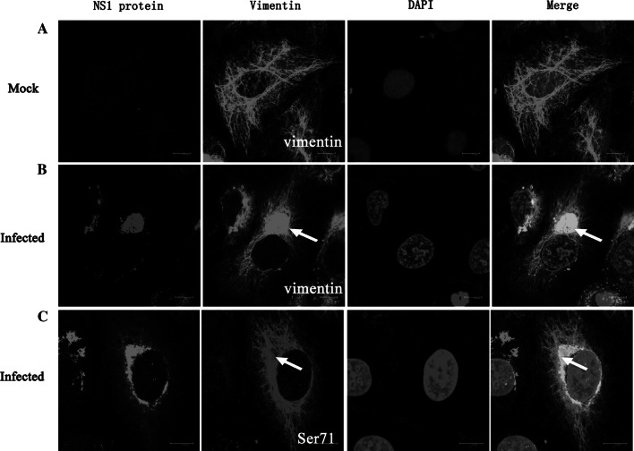
Fig. 2.
DENV2 infection induced aggregation of vimentin and Ser71-phosphorylated vimentin into dense structures at the perinuclear area. ECV304 cells were infected with DENV2 for 24 h. Immunostaining shows that the characteristic distribution of vimentin in control cells (a) completely changes in infected cells where vimentin (b) and phosphorylated vimentin (c) move to the perinuclear region and co-localize with DENV2 NS1 (arrows). Scale bars 10 μm

