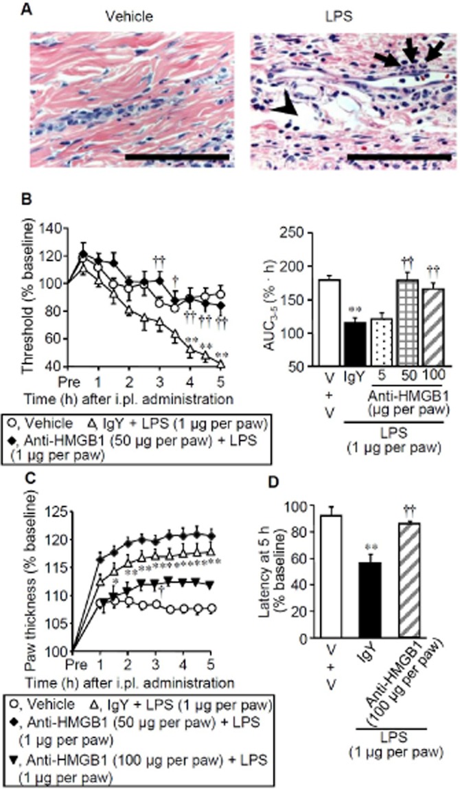
Figure 2.

Inflammatory hyperalgesia induced by LPS and its inhibition by the HMGB1-neutralizing antibody in rats. (A) Histopathological appearance of the hindpaw tissue 5 h after i.pl. administration of vehicle (left) or LPS at 1 μg per paw (right) in rats. The tissue was stained with haematoxylin-eosin. Arrows and arrow heads show slight infiltration of polynuclear leukocytes and oedematous lesion, respectively. Bars indicate 100 μm. (B–D) Mechanical nociceptive threshold (B), paw thickness (C) and paw withdrawal latency in response to thermal stimuli (D) after i.pl. combined administration of LPS and IgY or the HMGB1-neutralizing antibody in rats. Chicken anti-HMGB1 polyclonal antibody (neutralizing antibody) at 5, 50 or 100 μg per paw or chicken IgY at 50 μg per paw was co-administered i.pl. with LPS at 1 μg per paw. Area under the curve3–5 was calculated from the time-threshold curve between 3 and 5 h after i.pl. LPS (B, right), and the latency was determined 5 h i.pl. LPS (D). V, vehicle. Data show the mean with SEM for 4–10 rats. *P < 0.05, **P < 0.01 versus Vehicle; †P < 0.05, ††P < 0.01 versus IgY + LPS.
