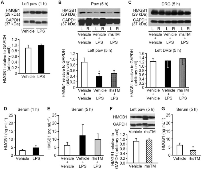
Figure 4.
HMGB1 protein levels in the hindpaw, DRG and serum after i.pl. administration of LPS in rats. rhsTM at 10 mg kg−1 or vehicle was administered i.p. 30 min before i.pl. administration of LPS at 1 μg per paw. (A–C) Expression of HMGB1 protein in the hindpaw plantar tissue 1 h (A) and 5 h (B) after i.pl. LPS, and in DRG at L4-L6 levels (C) 5 h after i.pl. LPS. The rats pretreated with rhsTM or not received i.pl. administration of LPS the left hindpaw, and HMGB1 levels in the left (L) and/or right (R) hindpaws (A,B) and in the left DRG (C) were assessed by Western blotting. (D,E) Serum HMGB1 levels 1 h (D) and 5 h (E) after i.pl. LPS in rats, as assessed by ELISA. (F,G) HMGB1 levels in the hindpaw (F) and serum (G) 5 h after i.p. administration of rhsTM at 10 mg kg−1 or vehicle in naïve rats. Photographs show typical examples of Western blotting for HMGB1 and GAPDH. Levels of each protein are quantified by densitometry in Western blotting. Data show the mean with SEM for 4 (A, B, C, D, F, G) or 5–8 (E) rats. *P < 0.05 versus vehicle + vehicle, or vehicle.

