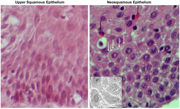
Fig. 1.
A light photomicrograph of a hematoxylin-eosin stained section of native esophageal upper squamous epithelium (left panel) and neosquamus epithelium from the lower esophagus (right panel). Note that neosquamous epithelium shows dilated intercellular spaces (white arrows) and has a prominent infiltrate of eosinophils (black arrows) when compared with the normal-appearing ‘native’ upper squamous epithelium. Magnification 60X. The INSERT in the right panel is an electron photomicrograph to better illustrate the dilated intercellular spaces in neosquamous epithelium. Magnification 3000X.

