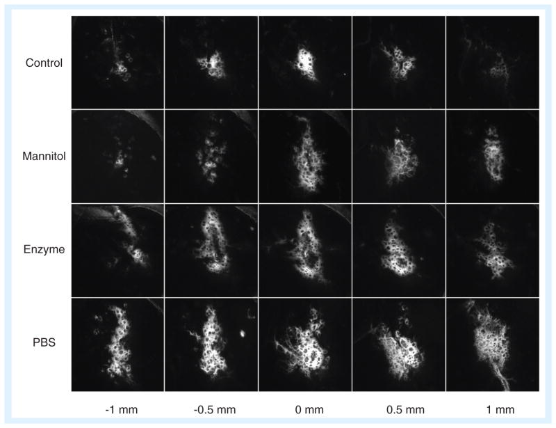
Figure 3. Nanoparticle transport in the brain.
Nanoparticles 30–40 nm in diameter were infused into the striatum of rats, either on their own in isotonic suspension (control), with hyperosmolar mannitol, or in isotonic suspension after pre-treatment with hyaluronidase or saline. The fluorescence image indicates the distribution of nanoparticles within 1 mm from the injection site. All three treatment conditions served to increase the effective pore size in the brain extracellular space during (mannitol) or prior to (enzyme, PBS) nanoparticle infusion, resulting in increased transport.
PBS: Phosphate-buffered saline.
Reproduced from [85] with permission from Elsevier.

