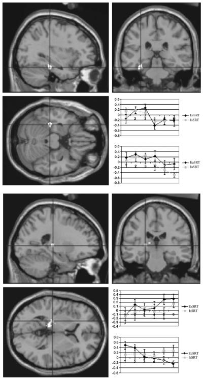
Figure 5.
Condition-specific differences in learning-related activation. Upper panel: comparison of EaSRT with IaSRT conditions. Left MTL shows an anttenuation of learning-related reductions in activation when subjects are explicitly trying to learn the sequence. The region showing this interaction is superimposed upon a representative MRI (x, y, z = −30, −30, −16). The inlaid plot shows averaged levels of activity in this region (relative to the fixation condition) for the standard SRT tasks (upper plot) and the alternating SRT (lower plot). The lower plot reflects the significant attenuation of learning-related decrease when explicit instructions are given. Lower panel: posterior thalamic differences in learning-related activation. Activation differences are shown for the direct comparison between EaSRT and IaSRT and are superimposed upon a standard MRI (x, y, z = −22, −24, 4). The accompanying plots shows a time-dependent decrease in activation for this regions during EaSRT. This effect was not seen in any of the other conditions, which showed trends in the opposite direction.

