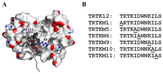
Figure 1.

Panel A The NMR solution structure of the wild-type TRTK12 peptide with rat S100B (PDB: 1MWN)(40). For clarity, the binding pocket of a single S100B monomer is shown. Key residues from the TRTK12 peptide are colored and labeled. Panel B: List of TRTK12 peptide sequences. Alanine-substitued residues are underlined.
