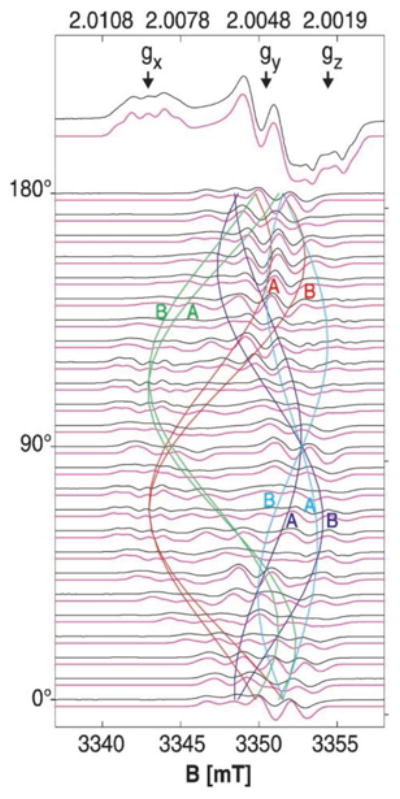
Figure 12.

These data show the single crystal rotation of the H2O2-induced tyrosine radical intermediate in ribonucleotide reductase. From the results, the researchers were able to determine the orientation of the tyrosine sidechain within the crystal, which is slightly rotated from X-ray diffraction structure of the resting state. Reproduced with permission from reference 127.
