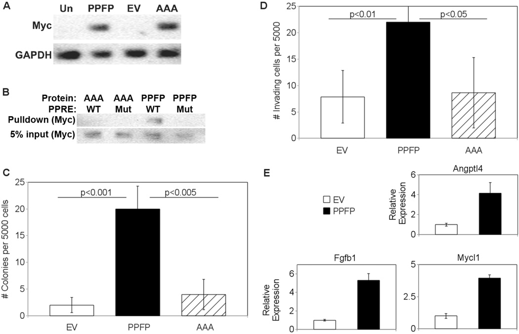
Figure 1.
(A) Expression of PPFP in stably transfected PCCL3 cells. Whole cell lysates were made from untransfected PCCL3 cells (Un), or PCCL3 cells stably transfected with either PPFP, empty vector (EV), or PPFP in which the P Box amino acids EGG were mutated to AAA (AAA). Twenty micrograms of protein per lysate were analyzed by Western blot for Myc, after which the blot was reprobed for GAPDH. PPFP is ~100 kDa and GAPDH is ~36 kDa. (B) P Box mutation AAA prevents binding of PPFP to a PPAR response element (PPRE). PCCL3-PPFP or PCCL3-AAA whole cell lysates were incubated with a biotinylated PPRE from the mouse Aqp7 gene (WT) or a mutated version (Mut), as described in Methods. Protein-DNA complexes were isolated with NeutrAvidin agarose beads and were analyzed for PPFP by Western blot using anti-Myc (top row). Five percent of input also was analyzed (bottom row). (C) Increased soft agar colony formation by PCCL3-PPFP cells. PCCL3-EV, -PPFP or -AAA cells were suspended in soft agar at 5000 cells per well and colony formation after 21 days was determined as described in Methods. Results are expressed as means ± SD and repeated with independent cell lines. (D) Increased invasion by PCCL3-PPFP cells. PCCL3-EV, -PPFP, or –AAA cells were plated on Matrigel-coated transwells with serum-free media and placed in tissue culture wells containing 10% FBS as attractant. After 36 hours, cells that invaded through the transwell membrane were counted under a microscope. Results are expressed as means ± SD and repeated with independent cell lines. (E) Upregulation in PCCL3-PPFP cells of genes overexpressed in human PPFP FTC. Five genes common to 2 published profiles of genes overexpressed in human PPFP FTC were analyzed by RT-qPCR in lysates from PCCL3-EV and PCCL3-PPFP cells. The 3 genes shown here had increased expression in the PCCL3-PPFP cells; the other two genes had no change (data not shown). Results are means ± SD.

