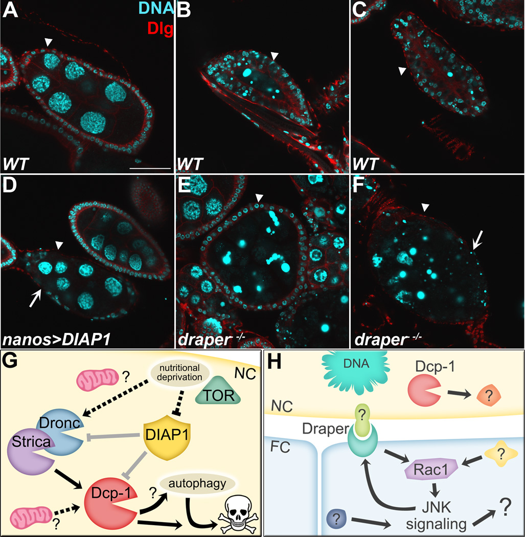
Figure 2. Overview of starvation induced cell death during mid-oogenesis.
Mid-stage egg chambers from starved flies stained with DAPI (cyan) to label DNA and anti-Discs large (Dlg, red) to mark the cell membranes. A–C) WT egg chambers. A) Healthy stage 8 egg chamber has large NC nuclei surrounded by a thin layer of FCs. Arrowhead indicates FC layer in all panels. B) Dying egg chamber has condensed and fragmented NC DNA, and the surrounding layer of FCs has enlarged and begun to engulf germline material. C) Late dying egg chamber has few NC nuclear fragments remaining and the FCs have completed engulfment. D) An undead egg chamber (arrowhead) where the NC nuclei have failed to condense and fragment, and many of the surrounding FCs have disappeared (arrow), resulting from overexpression of DIAP1 in the NCs. E) draper−/− mid-dying egg chamber contains a thin layer of FCs (arrowhead) that have failed to enlarge (compare to WT in B). F) Late dying draper−/− egg chamber (arrowhead) has lingering germline material and pyknotic FCs (arrow). Scale bar = 50 µm. G) Model of mid-stage death, showing suppression of caspases by DIAP1 (in gray) and potential regulatory mechanisms for mitochondria and nutritional deprivation (dotted lines). H) Model of mid-stage engulfment, showing activation of Draper-Rac1-JNK pathway by an unknown signal.

