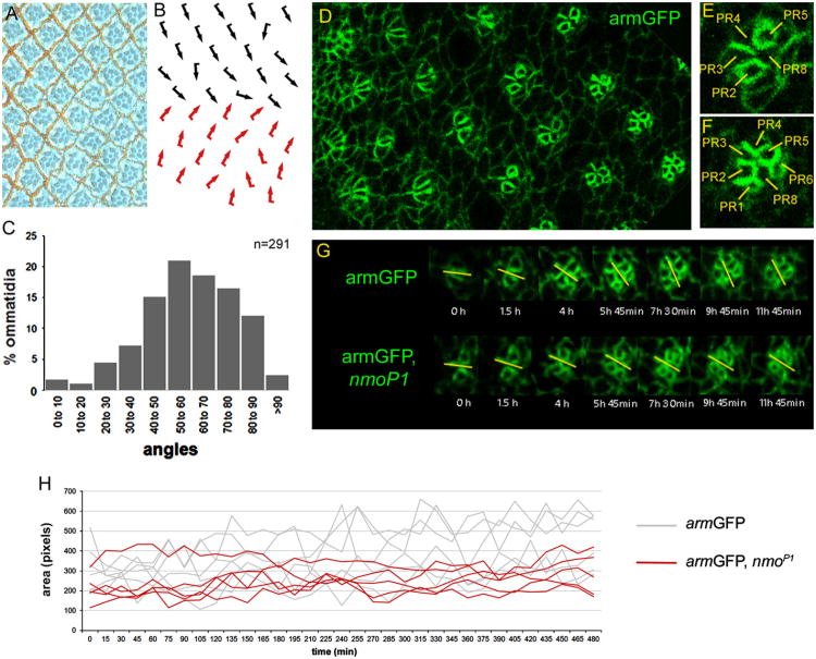
Fig. 1.
Live-imaging analyses in eye imaginal discs reveal that nmo regulates the rate of ommatidial rotation and IOCs dynamics. (A–B) Tangential section of armGFP, nmoP1 adult eye (A) and the corresponding schematic representation with ommatidia arranged around the equator (B), with dorsal and ventral chiral forms indicated by black and red arrows, respectively. (C) Bar chart illustrating the percentage of ommatidia (y-axis) that are oriented at the angles indicated (x-axis) in armGFP, nmoP1 eyes, in which the most represented angles range from 50° to 70°. (D-F) armGFP protein localization in eye imaginal discs. A transgene with the adherens junction protein linked to GFP labels apical cell contours and outlines cell boundaries in an area of the eye imaginal disc posterior to the morphogenetic furrow (D). Magnified views of an ommatidial precluster that has initiated rotation (E), in which the five photoreceptor (PR) cells are labeled with their numbers, and an older one (F), in which almost all the PRs have been recruited. (G) Time-lapse series showing individual ommatidia during rotation after ∼ 12 h from armGFP (upper panel) and armGFP, nmoP1 (lower panel) eye imaginal discs. The yellow bars mark the orientation angle of ommatidia with respect to the equator and the time on each photogram is referred to the first image of the series. The rotation rate of armGFP, nmoP1 ommatidia is slower than that of armGFP controls. (H) Quantification of several IOCs areas (number of pixels/cell) over time in armGFP (gray lines) and armGFP, nmoP1 (red lines) eye imaginal discs. Note that fluctuations of IOCs areas are sharper in wild-type controls than in nmo mutant discs, which is consistent with the observation that apical shape changes in IOCs are reduced in such mutants.

