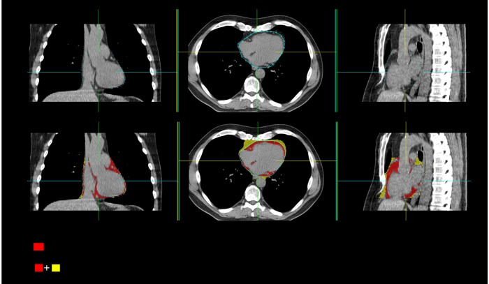
Figure 1.
Epicardial and thoracic fat measurement from non-contrast CT images acquired for coronary calcium scoring by a semi-automated method. Upper row: Pericardial sac is traced by placing control points on the pericardium. Lower row: Red overlay represents epicardial fat and yellow overlay represents extra-pericardial fat, together they represent total thoracic fat

