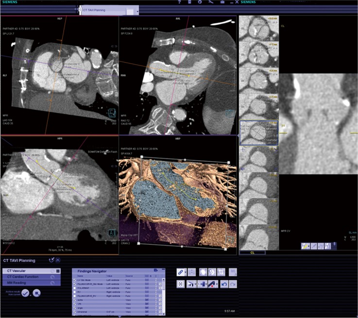
Figure 1.
LVOT and aortic root: using an advanced 3-D analysis program (syngo VIA, Siemens Medical Solutions), multiplanar reconstruction of the left ventricular outflow tract (LVOT) and aortic root is performed. The left sided panels show orthogonal MPR reconstructions of the aortic root, with the image plane focused on the annulus. A volume rendered image (VRI) image of the aortic root with the visible centerline is shown. The right sided panels show images reconstructed along and perpendicular to the centerline. Measurements are captured and saved as findings (bottom of the screen)

