Figure 5.

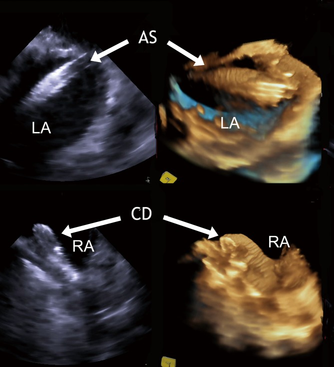
RT-3D ICE: comparative imaging of a access sheath passing a PFO (upper panel) and a fully deployed PFO device (lower panel) by 2D and RT-3D ICE. LA, left atrium; RA, right atrium; ES, access sheath; CD, closure device

RT-3D ICE: comparative imaging of a access sheath passing a PFO (upper panel) and a fully deployed PFO device (lower panel) by 2D and RT-3D ICE. LA, left atrium; RA, right atrium; ES, access sheath; CD, closure device