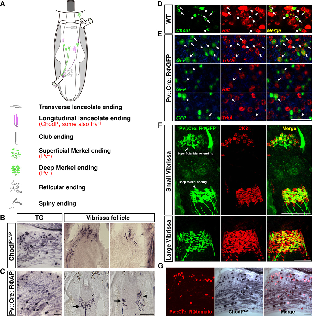
Fig. 1. Genetic labeling of RA-longitudinal lanceolate and SA-Merkel ending neurons in the vibrissa sensory system.
(A) Schematic drawing of the types of touch sensory endings in the mouse vibrissa follicle.
(B) Representative images of AP staining result from P7 ChodlPLAP/+ mice on trigeminal ganglion (TG) section (left) and vibrissa follicle sections (right two panels). Longitudinal-lanceolate-ending neurons were selectively labeled.
(C) Representative images of AP staining result from P7 Pv::Cre; RΦAP mice on TG (left) or vibrissa follicle sections (right two panels). Pv::Cre; RΦAP labeled Merkel-ending (arrow) and a few longitudinal lanceolate-ending (arrowhead) neurons.
(D) Representative images of 2-color fluorescent in situ hybridization with Chodl (green) and Ret (red) probes on the P7 WT TG sections. 96.46 ± 0.27% chodl+ cells co-expressed Ret (arrow).
(E) Representative images of 2-color fluorescent in situ hybridization on TG sections from P21 Pv::Cre; RΦGFP mice. Top row: GFP (green) and TrkC (red); middle row: GFP and Ret; bottom row: GFP and TrkA. 62.4 ± 1.5% GFP+ cells (labeled by Pv::Cre) co-expressed with TrkC. 25.6 ± 1.0% GFP+ cells co-expressed with Ret. 17.3 ± 2.9% GFP+ cells co-expressed with TrkA.
(F) Double immunostaining showing sensory axons (green) and Merkel cells (red) on sections from the mystacial pad from Pv::Cre; RΦGFP mice at P7. In the small vibrissa (top row), more than 90% of Merkel cells were innervated by Pv::Cre labeled axons; in contrast, in the large vibrissa (bottom row images), Pv::Cre only labeled subsets of axons innervating Merkel cells.
(G) Representative TG sections from Pv::Cre; RΦtomato; ChodlPLAP/+ triple transgenic mice (P7). 22.3 ± 0.8% Chodl+ (AP stained) neurons exhibit tomato fluorescence (PLAP and tomato double positive). Conversely, 24.3 ± 1.3% tomato+ neurons exhibit PLAP activity (Chodl+).
Scale bars: 100 µm.
See also Figure S1.

