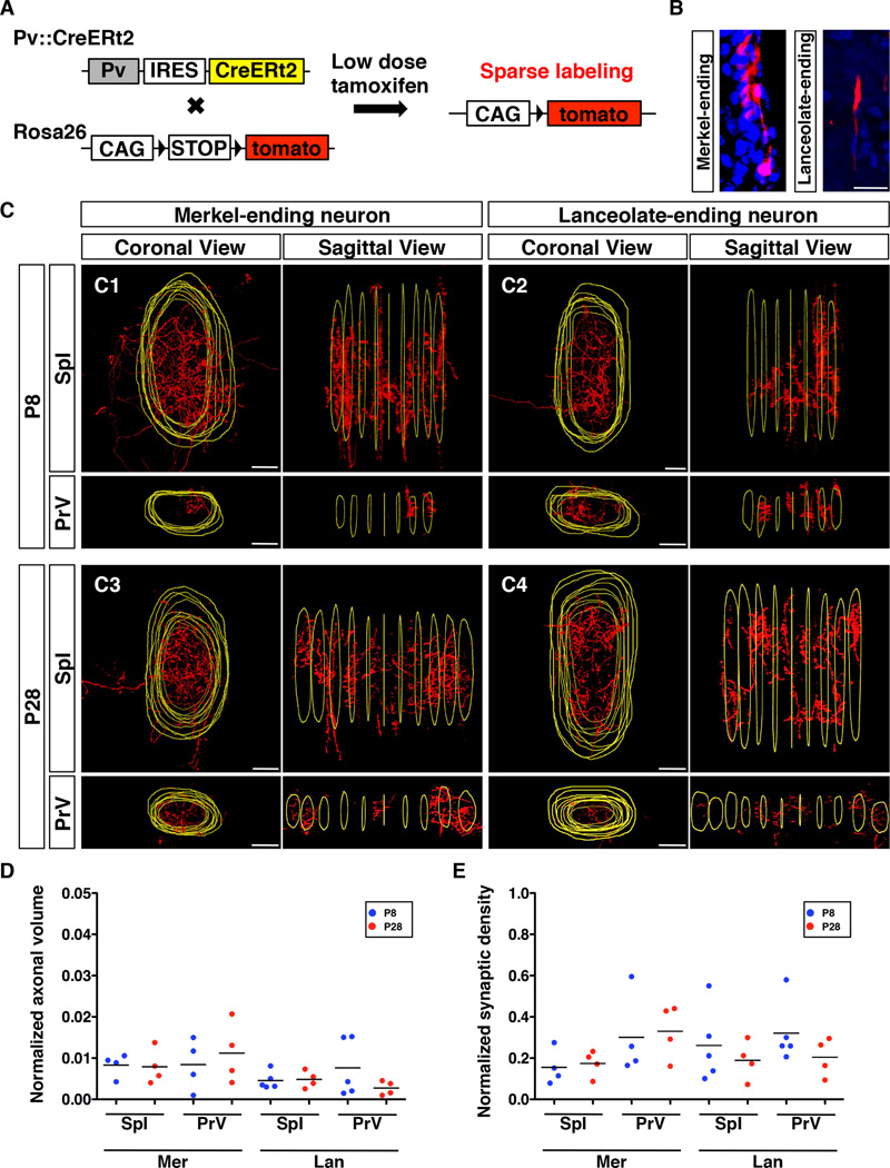
Fig. 3. Axon collaterals of single-labeled RA-lanceolate and SA-Merkel neurons inside the barrelette unit.
(A) Schematic representation of the strategy used to sparsely label longitudinal lanceolate and Merkel-ending neurons.
(B) Representative images of the peripheral ending of single-labeled Merkel or lanceolate neuron. Red, axon; Blue, DAPI.
(C) Representative coronal views and lateral/sagittal views of 3D reconstructed collaterals from single-labeled Merkel and single-labeled lanceolate neuron in the SpI and PrV at P8 and P28, respectively. Red, axon; Yellow, outlines of the barrelette structure.
(D) Quantification of relative volumes occupied by axon collaterals of single-labeled Merkel or lanceolate neurons normalized with respect to the volume of each corresponding barrelette column. All comparisons between P8 and P28 are not statistically different (p>0.05, t-test).
(E) Quantification of relative synaptic densities of single-labeled Merkel or lanceolate neurons normalized with respect to the total volume of the collaterals. All comparisons between P8 and P28 are not statistically different (p>0.05, t-test).
Abbreviations: Mer, Merkel-ending neuron; Lan, lanceolate-ending neuron. Scale bars: 20 µm.

