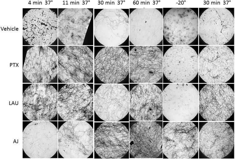
Figure 3.

Electron microscopy of microtubules. Electron micrograph images of microtubules formed in the presence of 2 mg/ml tubulin with 10 μM PTX, LAU, AJ or vehicle 4, 11, 30 or 60 min after tubulin reactions were warmed to 37°C, after chilling the reactions at -20°C for 30 min or after repolymerization at 37°C for 30 min after chilling. All images were acquired at a magnification of 2,000×.
