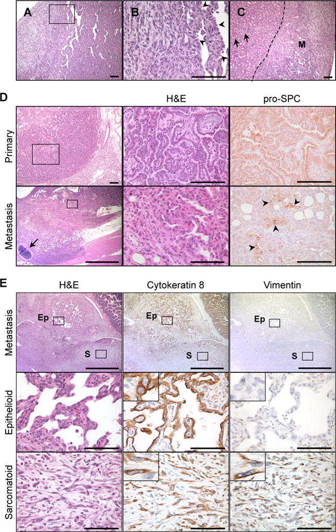
Figure 2. Poorly differentiated, sarcomatoid lung tumors develop in Arf-deficient animals.

[A] Pulmonary sarcomatoid carcinoma-like lesions formed in the lung. [B] Magnification of boxed region in [A]. Arrowheads point to well-differentiated, epithelial regions. [C] Renal metastasis (M) of mixed epithelial and sarcomatoid components. Arrows indicate normal kidney glomeruli. [D] Lung adenocarcinomas (top) and sarcomatoid metastases (bottom) from the same mouse exhibited immunoreactivity for pro-surfactant protein C (pro-SPC), a marker of lung epithelial cells. Bottom panel displays invasion of the pleura and chest wall. Arrow indicates rib. The metastatic tumor maintained regions of epithelial differentiation (boxed region, magnified in middle panel) that expressed pro-SPC (arrowheads). [E] Metastasis of sarcomatoid carcinoma to peritoneal cavity. Whereas both the sarcomatoid (box S) and epithelioid (box Ep) compartments were positive for cytokeratin 8, vimentin expression was restricted to the sarcomatoid regions. Scale bars 100 μm, except panel [D, bottom left] (1 mm).
