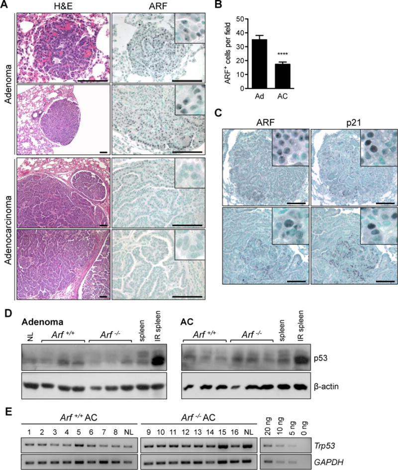
Figure 4. The ARF-p53 signaling pathway.

[A] Adenomas (Ad) from Arf+/+ mice robustly expressed ARF, but adenocarcinomas (AC) frequently lost ARF expression. Inset shows nucleolar staining of ARF in adenomas. [B] Quantification of ARF positive cells (**** P < 0.0001; n = 46 Ad and 145 AC fields examined). [C] ARF and p21 proteins colocalize by IHC in adenomas (top) and regions of low-grade adenocarcinomas (bottom) in wild-type mice. All scale bars 100 μm. [D] Western blot analysis of p53 reveals expression of p53 protein in adenomas and adenocarcinomas isolated from both genotypes. Irradiated spleen is positive control. β-actin is provided as loading control. [E] PCR amplification of genomic DNA shows that the Trp53 locus has not been deleted in adenocarcinomas from either genotype. GAPDH is provided as loading control. A dilution series is included to demonstrate that the PCR was performed in the exponential range. NL = normal lung.
