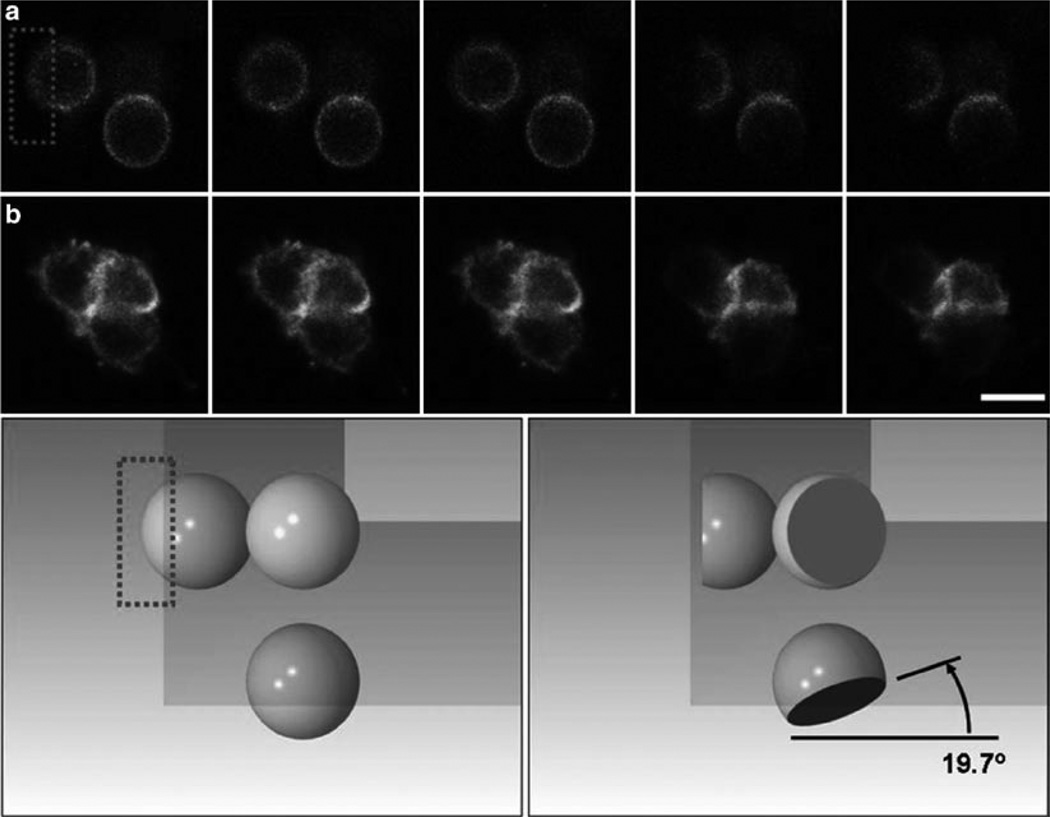
Fig. 6.
Fluorescence recovery after photobleaching (FRAP) experiments in mirrored pyramidal wells (MPWs). Shown is a series of two individual cells expressing cAR1-GFP along with their reflections prior to and after selective photobleaching in an MPW. Top panel: Five successive frames of a Latrunculin-treated, immobilized cell (top row, a) and an untreated cell (bottom row, b), each of which was positioned in the corner of an inverted MPW. In each case, laser scanning of the upper left reflected image leads to selective photobleaching of the dorsal surface of the cell, as can be seen in reflected images in the same frame (bleaching occurs after frame 3 in each series). The angle of the photobleached section is very near the expected angle. Bottom panel: A SolidWorks simulation of the photobleach experiment illustrating the cell before bleaching (left) and after bleaching (right), and the shape of the bleached section of the sphere as seen from above and from both reflections. Image is courtesy of Kevin Seale. Bar is 15 µm.

