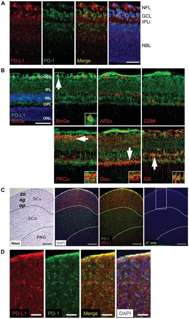
FIG. 1.
PD-L1 expression in the neonatal mouse retina and brain by immunofluorescence staining. A. PD-1 (green) and PD-L1 (red) expression at P2 in the central retina, at the level of the optic nerve. B. PD-L1 (green) and the following retinal cell type markers (all in red): Brn3a for RGCs, AP2α for amacrine cells, Calbindin D28K (D28K) for horizontal cells, PKCα for rod bipolar cells, Goα for optic nerve bipolar cells, glutamine synthetase (GS) for Müller glia. White arrows indicate cells shown at higher magnification inset into each panel. All images were taken at the level of the optic nerve, at the midpoint between the optic nerve head (ONH) and periphery. C. Coronal section of the superior colliculus at Bregma −4.3 mm, after Allen Mouse Brain Atlas coordinates. Panels from left to right show: 1) Nissl stain, 2) PD-1 (green) and PD-L1 (red) expression by immunofluorescence, with DAPI counterstain (blue), 3) same as second image, without DAPI, and 4) secondary only negative control, where PD-1 and PD-L1 primary antibodies were omitted, and the white box indicates area shown at higher magnification in D. D. Higher magnification of SCs. Panels from left to right show: 1) PD-L1 (red) expression, 2) PD-1 (green) expression, 3) PD-L1 and PD-1 expression colocalization (yellow), and 4) PD-L1 and PD-1 expression with DAPI (blue) counterstain. Scale bars = 50 μm (A, B, D) and 200 μm (C). For each expression pattern shown, 3 animals were examined. Nuclei were visualized with DAPI (blue) counterstain. Yellow represents colocalization of PD-1 and PD-L1. GCL, ganglion cell layer; INL, inner nuclear layer; IPL, inner plexiform layer; IPLi, incipient inner plexiform layer; NBL, neuroblast layer; NFL, nerve fiber layer; ONL, outer nuclear layer; OPL, outer plexiform layer; op, stratum opticum; PAG, periaqueductal gray; SCs, superior colliculus superficial layers; SCd, superior colliculus, deep layers; sg, stratum griseum superficiale; zo, stratum zonale.

