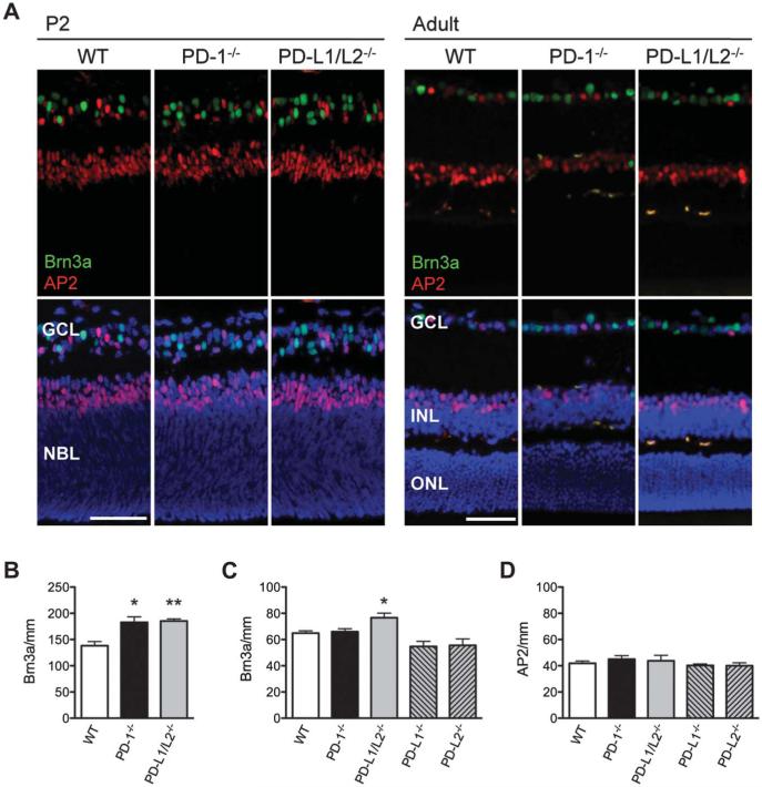
FIG. 2.
Increased RGCs in the neonatal and adult retina of PD-L1/L2−/− mice. A. Immunofluorescence staining for Brn3a (green) and AP2α (red) in P2 and adult (P56) retina of WT, PD-1−/−, and PD-L1/L2−/− mice, with DAPI nuclear counterstain (blue). Scale bars = 50 μm. B. Quantification of RGC number (Brn3a/mm) in WT, PD-1−/− mice at P2. Quantification of RGC number (Brn3a/mm, B, C) and amacrine cell number (AP2/mm, D) in the adult retina. Three mice per strain were analyzed at each time point. For each quantified readout, the mean and standard error of the mean were plotted. *Significant P value in comparing each knockout to WT; *P ≤ 0.04, **P = 0.006.

