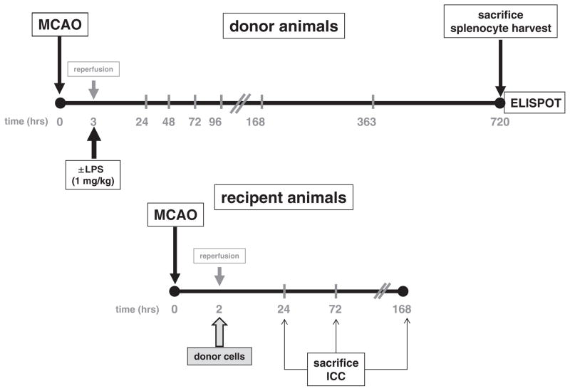
Fig. 1. Experimental protocol.
Donor animals underwent 3 h of MCAO (or sham surgery) and received either LPS or saline at the time of reperfusion. Animals were sacrificed at 720 h (1 month), splenocytes harvested and cells cultured with MBP for 48 h. A subset of cells was subjected to ELISPOT assay to determine the Th1 and Th17 responses to MBP. Recipient animals underwent 2 h MCAO and donor cells were injected into recipient animals at the time of reperfusion. Neurologic function was assessed at 24 h (1 day), 72 h (3 days) and 168 h (1 week) after MCAO. Subsets of animals were sacrificed at each time point for histological analysis.

