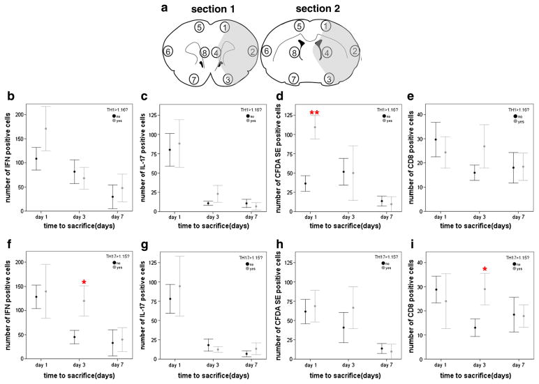
Fig. 3.
The numbers of cells staining for IFN-γ, IL-17, fluorescein and CD8 were determined in 4 regions within the infarcted hemisphere and in 4 regions within the non-infarcted hemisphere in two different coronal brain sections (a). Cells were counted in 6 adjacent high power fields (100×) within each of the 4 brain regions in the infarcted and non-infarcted hemispheres. The graphs show the total cell counts for regions 1 through 4 in both coronal sections (as there were virtually no identifiable cells in non-infarcted hemisphere). Animals receiving cells from a Th1(+) donor had more CFDA SE positive (or fluorescein+) cells in the infarcted hemisphere at day 1 after MCAO (d), but there were no differences in the number of IFN-γ+ (b), IL-17+ (c) or CD8+ (d) cells. Animals receiving cells from a Th17(+) donor had more IFN-γ+ and CD8+ cells at day 3 after MCAO (f and i). Data are presented as the mean and SEM. *P < 0.05 and **P < 0.01 by t-test.

