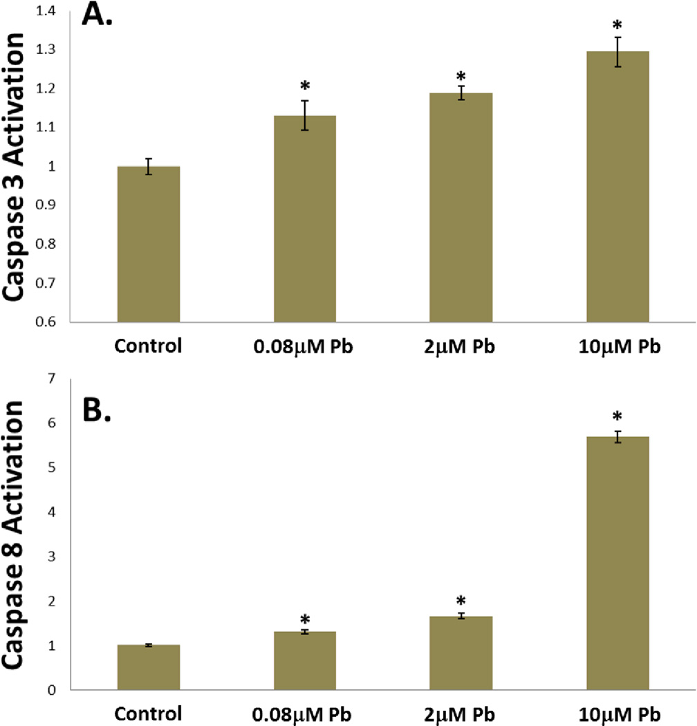
Figure 5.
Lead treatment resulted in dose-dependent caspase activation. As evidenced by high-specificity fluorescent indicator dyes, after 24hrs of lead treatment caspase3 activation was elevated approximately 15% at 0.08µM and significantly increased by 20% at 2µM and 30% at 10µM in chick articular chondrocytes (A.). Similarly, when treated with lead for 24hrs, caspase8 activation was increased in a dose-dependent manner, with a 6 fold increase seen in the 10µM treatment group (B.). (*=p<0.05 from control, n=8).

