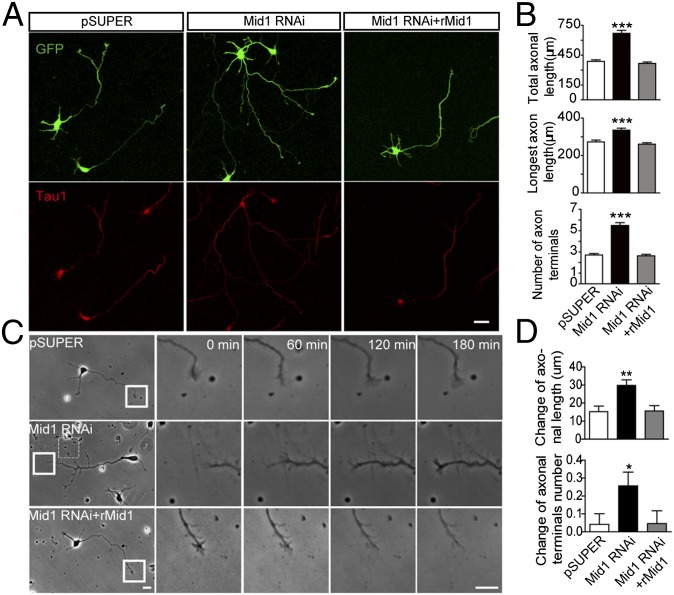
Fig. 2.
Knockdown of Mid1 promotes axon growth and branching in vitro. (A) Representative images of neurons transfected with GFP and the constructs indicated. At 4 DIV, the neurons were stained for GFP and Tau1 to visualize the morphology of whole cells and axons. (B) Quantification of total axonal length, longest axon length, and number of axonal terminals in neurons transfected with pSUPER, Mid1 RNAi, and Mid1 RNAi plus rMid1. More than 100 neurons from four independent experiments were analyzed in each group; data are shown as mean ± SEM, ***P < 0.001, Student t test. (C) Dynamics of axonal tips. Neurons transfected with the plasmids indicated were imaged for 180 min at 2 DIV. Right are higher magnification images of the boxed regions at the time points indicated. Magnified images of the dashed box region are shown in Fig. S2F. (D) Quantification of neurite dynamics. The change of axon length and number of axon terminals over 180 min was measured and shown as mean ± SEM, n = 48 in pSUPER group, n = 74 in Mid1 RNAi group, n = 87 in Mid1 RNAi+rMid1 group, *P < 0.05, **P < 0.01, t test. (Scale bars: 20 μm.)

