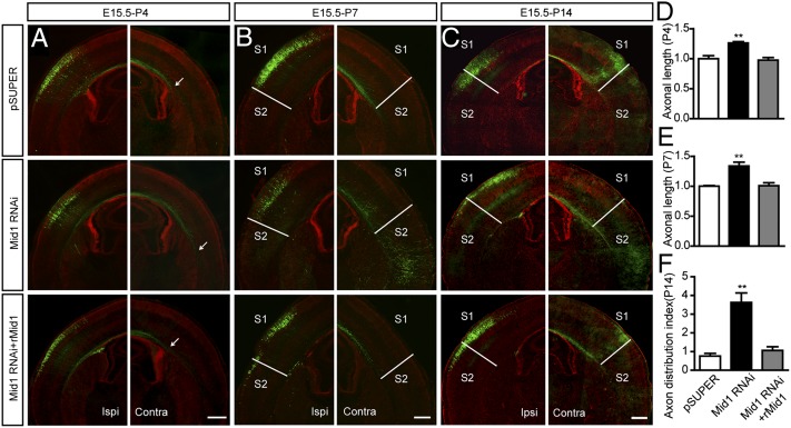
Fig. 3.
Silencing Mid1 accelerates callosal axons growth and changes their projection pattern. (A–C) The constructs pSUPER, Mid1 RNAi, or Mid1 RNAi plus rMid1 were coelectroporated with GFP into the ventricles of E15 mice. Animals were killed at different developmental stages. Brain slices at the level of Bregma −1.58 mm were stained with Hoechst and GFP antibodies to visualize the callosal axons. Arrows indicate the location of axon terminals. The S1/S2 border is labeled with white lines. (Scale bar: 500 μm.) Contra, contralateral side to electroporation; Ipsi, ipsilateral side to electroporation; S1, primary somatosensory cortex; S2, second somatosensory cortex. (D–F) Quantitative analysis of callosal axon length and axon distribution index. Results are shown as mean ± SEM. At P4, n = 4 in each group; at P7 and P14, n = 5–7 in each group. **P < 0.01, Student t test.

