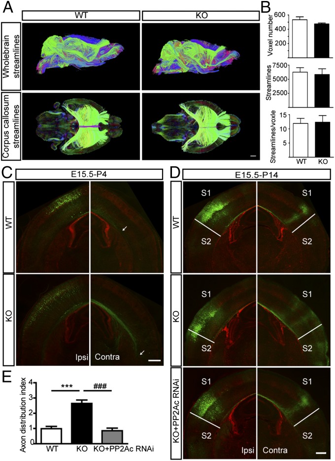
Fig. 5.
Corpus callosum development is abnormal in Mid1 KO mice. (A) Comparison of the corpus callosum (CC) between Mid1 KO and WT adult mice using DTI. (Upper) Representative whole-brain streamlines in a lateral-sagittal view of WT and KO mice. (Lower) CC streamlines in a dorsal view of WT and KO mice. Streamline color follows the following orientation code: green, mediolateral; red, rostrocaudal; blue, dorsoventral. (B) The volume of the CC was measured by voxel number, the number of streamlines, and the number of streamlines per voxel. n = 3 in each group. (C) Callosal axon growth is accelerated in Mid1 KO mice. The GFP plasmid was electroporated into neuronal progenitors at E15, and GFP staining was performed in brain slices of P4 animals. Arrows indicate the axon terminals within the corpus callosum. (D) Callosal projections are abnormal in Mid1 KO mice. GFP or PP2Ac RNAi constructs were electroporated at E15. Brain slices (Bregma −1.58 mm) from P14 Mid1 KO mice and their WT littermates were stained for GFP to visualize the callosal projection. (E) Statistical analysis of axon distribution index in D. Results are shown as mean ± SEM, n = 7–8 in each group. ***P < 0.001, compared with WT; ###P < 0.001, compared with KO; Student t test. (Scale bars: A, 1 mm; C and D, 500 μm.)

